Pregnancy Ultrasound in Pitampura: A Comprehensive Guide by Dr. Anuj Thakral
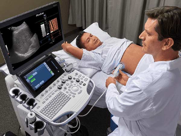
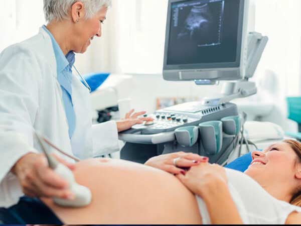
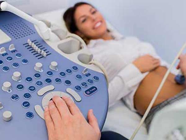
Pregnancy ultrasound is a pivotal aspect of prenatal care, offering invaluable insights into the development and well-being of the fetus. In Delhi, Dr. Anuj Thakral stands out as a leading expert for Level 1 & 2 pregnancy ultrasound in Pitampura, providing exemplary services to expectant mothers. Understanding the different levels of pregnancy ultrasound can empower individuals with the knowledge they need throughout their journey to parenthood.
Introduction to Pregnancy Ultrasound
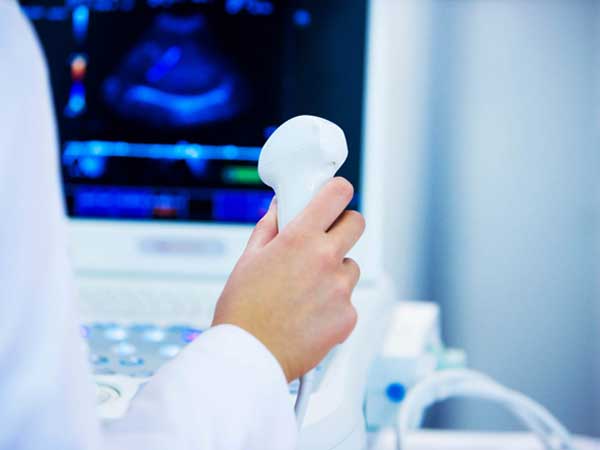
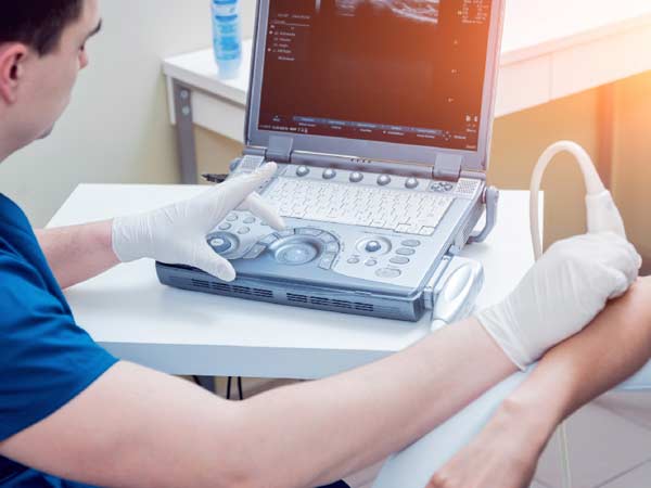
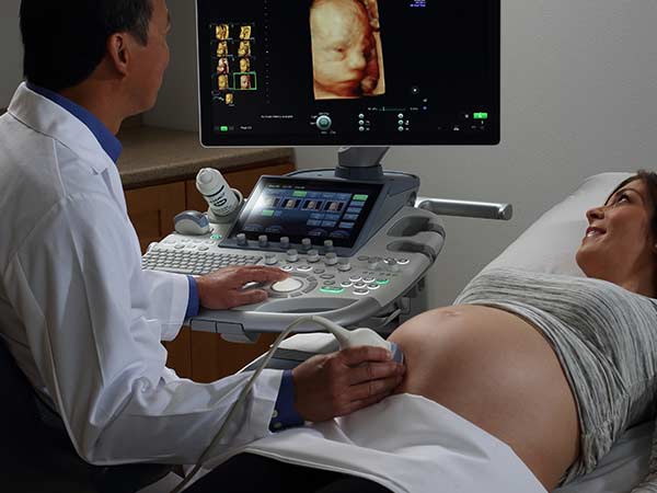
Pregnancy ultrasound, also known as sonography, is a non-invasive imaging technique used to monitor the progression of pregnancy. By utilizing sound waves, it provides detailed images of the fetus, placenta, and uterus, allowing healthcare providers to assess fetal development and detect any potential complications.
Importance of Pregnancy Ultrasound
Ultrasound plays a crucial role in prenatal care by enabling healthcare providers to:
- Monitor fetal growth and development.
- Assess the placenta and amniotic fluid levels.
- Identify any abnormalities or birth defects.
- Determine the baby’s position and estimate its size.
- Evaluate the health of the mother’s reproductive organs.
Why choose Dr. Thakral’s Diagnostic Centre for level 1 & 2 pregnancy ultrasound in Pitampura
When considering where to go for level 1 & 2 pregnancy ultrasounds in Pitampura, there are several reasons why Thakral’s Diagnostic Centre stands out as an excellent choice:
- Expertise: Dr.Thakral’s Diagnostic Centre boasts a team of highly skilled and experienced radiologists and sonographers who specialize in prenatal imaging. With their expertise, you can trust that your ultrasound will be conducted with precision and accuracy.
- State-of-the-art Equipment: The centre is equipped with advanced ultrasound machines that produce high-quality images, allowing for detailed assessments of fetal development at every stage of pregnancy. This ensures that any potential abnormalities or concerns can be identified early on.
- Comprehensive Services: Whether you need a basic ultrasound to confirm your pregnancy or a more detailed level 1 ultrasound for specialized assessment, Dr.Thakral’s Diagnostic Centre offers a full range of prenatal imaging services to meet your needs. They also provide level 2 ultrasounds for comprehensive anatomical scans.
- Patient-Centered Care: At Dr. Thakral’s Diagnostic Centre, patient care is paramount. The staff are dedicated to providing compassionate and personalized care to every expectant parent who walks through their doors. They understand the importance of this moment in your life and strive to make your ultrasound experience as comfortable and stress-free as possible.
- Convenient Location: Situated in Pitampura, Thakral Diagnostic Centre offers a convenient location for residents in the area. Whether you live nearby or are traveling from elsewhere in Delhi, you can easily access their facilities for your ultrasound appointments.
- Prompt and Efficient Service: Time is of the essence when it comes to prenatal care. Thakral Diagnostic Centre is known for its prompt and efficient service, ensuring that you receive your ultrasound results in a timely manner. This allows for early detection of any issues and timely interventions if necessary.
What are the types of Ultrasound studies in pregnancy (trimester wise):
First-trimester ultrasound

There are generally two ultrasounds offered during the first 3 months (First trimester) of pregnancy:
- Early pregnancy scan or viablity scan (At 6-8 weeks of pregnancy): Helps to confirm and determine pregnancy age, hence the name dating scan. This pregnancy scan can detect the baby’s heartbeats and any source of internal bleeding or abnormality found in the uterus that might affect the pregnancy.It also confirms the location of pregnancy and checks if it is inside or outside the uterus. Pregnancies outside the uterus are called ectopic pregnancies. Such pregnancies need early diagnosis and immediate medical attention. Twin pregnancy or multiple pregnancies can also be detected at this stage.
The dating scan is very important as it establishes a benchmark which will enable accurate assessment of fetal growth as the pregnancy progresses in the 2nd & 3rd trimester.
2. Nuchal Translucency Scan (NT/NB ultrasound pregnancy scan)
(11 week to 13 week 6 days ultrasound scan): Most suitable time is 12-13 weeks
“Nuchal” is the adjective for the “nape of the neck”, and translucency here refers to the translucent area in that portion. The fluid at the back of your baby’s neck is checked by this scan. The NT scan looks at the entire spinal column of the baby, measures the size and thickness of its nuchal translucency. NT is increased in up to 40% of fetuses that have a major cardiac abnormality and is associated with other structural and genetic anomalies.
This scan also checks for the presence of nasal bone.The nasal bone is ‘absent’ or hypoplastic in 50–60% of fetuses with trisomy 21.
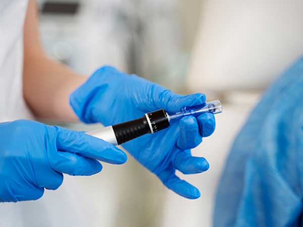
Alongwith this sonography, a double marker blood test is recommended. This sonography and double marker blood test guides obstetricians to rule out chromosomal abnormalities like Down’s syndrome.
We routinely perform uterine artery doppler as part of NT/NB scan. This helps us prempt possiblity of increased blood pressure and related complications in the mother and growth issues in baby in later pregnancy.
Second-trimester ultrasound

Level 2 Pregnancy Ultrasound: A Detailed Look
Reasons for Level 2 Ultrasound
A Level 2 ultrasound, also known as an anatomy scan or targeted ultrasound, provides a more comprehensive evaluation of fetal anatomy and development. It is typically performed between 18-22 weeks of pregnancy.
What’s Examined
During a Level 2 ultrasound, Dr Anuj Thakral examines the fetus in detail, focusing on the brain, heart, spine, limbs, and other vital organs. This scan can detect structural abnormalities and assess the baby’s overall health.
When it’s Done
The timing of a Level 2 ultrasound is critical, as it allows Dr Anuj Thakral to detect any anomalies early in the pregnancy. Early detection can lead to timely interventions and better outcomes for both the mother and baby.
3. Anomaly scan/TIFFA scan (Level 2 scan)- at 18 to 20 weeks
An anomaly scan or TIFFA scan (Targeted Imaging for Fetal Anomalies) is the most essential pregnancy scan carried out in your second trimester.
The anomaly scan assesses the baby’s structural anatomy & internal organs from head to toe in adequate detail. Besides recording the fetal size & weight, a detailed anomaly scan can help in timely detection of a variety of major congenital anomalies/malformations. For this, special attention is paid to the brain, face, spine, heart, stomach, bowel, kidneys, and limbs..
During the anomaly scan, we also assess the position of placenta, the umbilical cord, and amniotic fluid around the fetus. A uterine artery doppler is also an essential part of the study.

Fetal Echocardiography
This ultrasound is meant to check the structure and function of a baby’s heart.
This is one of the optional sonographies during pregnancy. This pregnancy ultrasound scan is recommended typically in high-risk pregnancies. It’s typically done in the second trimester, between weeks 18 to 24.
This scan looks out for abnormalities such as holes in the heart, narrowing of arteries, valves that don’t open and close properly. The common indications for fetal echocardiography include:
- Mother having a heart defect
- Mother already has had a baby with a heart defect
- Diabetic mother: a slightly higher risk of having a baby with a heart defect
- History of consuming certain drugs that can increase the risk of heart problems, such as some antiepileptic medications
- Abnormal nuchal translucency (NT) scan with normal chromosomal study

Third-trimester ultrasound

In the last 3 months of pregnancy, ultrasound is used to monitor the baby’s health, growth, development, and internal organs. Changes in the uterine environment are also carefully assessed. Following are the pregnancy ultrasound scan prescribed in the third trimester :
- Third Trimester growth & Fetal well being scan: besides fetal growth & anomalies, the position of placenta & amount of amniotic fluid is assessed. The lower end of the placenta should not be lying too close to the cervix. In the case of twins or triplets, there is a greater chance of developing growth problems, which is checked by the growth scan.
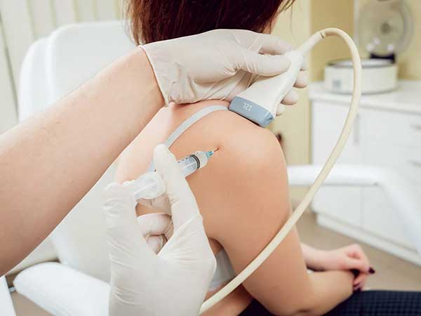
- Obstetric Doppler scan: A proper blood flow to the uterus, placenta, and the umbilical cord is crucial for a baby to develop normally inside the womb during pregnancy.
A routine pregnancy ultrasound alone is incapable of demonstrating the blood flow. It is complimented by color doppler which looks at both the maternal vessels (uterine arteries) & fetal vessels (umbilical & middle cerebral arteries in most cases). This helps identify & quantify deficencies in maternal & fetal blood flow which are important to guide further management in this final trimester.
Benefits of Early Detection
Early detection of fetal abnormalities or complications allows healthcare providers to develop a comprehensive care plan and provide appropriate support and interventions to ensure the best possible outcomes for both the mother and baby.
Risks and Safety Concerns
While ultrasound is considered safe for both the mother and fetus, it is essential to minimize unnecessary exposure to ultrasound waves. Dr Anuj Thakral follows strict guidelines to ensure the safety and well-being of both patients during ultrasound procedures.
Choosing the Right Ultrasound Facility
When selecting an ultrasound centre in Pitampura, it is essential to consider factors such as the expertise of the healthcare providers, the quality of the equipment, and the availability of comprehensive services tailored to individual needs.
Understanding the Role of Dr. Anuj Thakral
Dr. Anuj Thakral is a renowned radiologist specializing in level 1, 2, & 3 pregnancy ultrasound in Pitampura. With his expertise and compassionate care, he has helped numerous families navigate the journey of pregnancy with confidence and peace of mind.
Tips for a Successful Ultrasound Experience
- Schedule regular prenatal appointments to monitor the progress of your pregnancy.
- Stay hydrated and follow any specific instructions provided by your healthcare provider before the ultrasound.
- Communicate openly with your healthcare provider about any concerns or questions you may have.
- Take time to bond with your baby during the ultrasound by asking questions and requesting to see images of your little one.
Conclusion
Pregnancy ultrasound plays a pivotal role in prenatal care, offering invaluable insights into fetal development and well-being. By understanding the different levels of pregnancy ultrasound and the expertise of Dr Anuj Thakral, expectant parents can embark on their journey to parenthood with confidence and peace of mind.
Overall, Thakral Diagnostic Centre is a trusted choice for level 1, 2, and 3 pregnancy ultrasounds in Pitampura, offering expertise, advanced technology, comprehensive services, patient-centered care, and convenience all under one roof.
Frequently Asked Questions (FAQs)
Is ultrasound safe during pregnancy?
Yes, ultrasound is considered safe for both the mother and fetus when performed by trained healthcare professionals following established guidelines.
How often should I have a pregnancy ultrasound?
The frequency of ultrasound exams may vary depending on your specific healthcare needs and any underlying medical conditions. Your healthcare provider will determine the appropriate schedule for you.
Can ultrasound detect all birth defects?
While ultrasound is an essential tool for detecting many birth defects, it cannot identify every possible anomaly. In some cases, additional tests or screenings may be necessary.
What should I expect during a pregnancy ultrasound?
During a pregnancy ultrasound, you will lie on an examination table while a transducer is moved over your abdomen to capture images of your uterus and fetus. The procedure is painless and typically takes around 20-30 minutes.
How can I prepare for a pregnancy ultrasound?
Your healthcare provider will provide specific instructions on how to prepare for your ultrasound, which may include staying hydrated and avoiding urinating before the exam.
List of documents/information needed for any ultrasound done on a pregnant women (as per guidelines of PC/PNDT Act):
- Identity proof of patient having full name (eg. Aadhar card) matching the prescription
- Date of first day of last menstrual period (LMP) of pregnant lady
- Proper prescription by referring doctor mentioning indication for ultrasound- valid only for 21 days from date of issue
- Prescription to have full name of referring doctor
- Prescription to have proper stamp of referring doctor with DMC number