Category Archives: Services

Ultrasound Guided Interventions

Ultrasound-guided interventions

Technology is bringing more and more facilities to our daily lives. In the field of medicine, ultrasound-guided procedures ensure that some diagnostic & therapeutic procedures can be performed with minimal invasion of the body.

Created in the First World War to detect objects at the bottom of the sea, ultrasound only arrived in medicine in the 1940s, when it revolutionized diagnostic procedures – even with the patient having to be submerged in a bathtub and the quality of the images being very low.

Nowadays, the quality of images has reached levels of extreme quality, and allow procedures guided by ultrasound to reach a level of excellence.

ULTRASOUND-GUIDED PROCEDURES

Among interventional medicine, ultrasound has also served to guide physicians in minimally invasive procedures. Through the images, it is possible to have more precision of the place where it is necessary to carry out interventions, such as knowing the exact place & path where it is necessary to insert a needle. Hence, a variety of Interventional Ultrasound guided procedures can be performed which can include the following:.

  • Ultrasound guided Fine Needle Aspiration Cytology (FNAC)– using a thin sterile metallic needle placed after local infiltration of anaesthetic agent. Needle targetted to nodule/mass/abnormality visualised on focussed ultrasound. Most useful for relatively vascular organs like Thyroid & targets like small lymphnodes where biopsy may have risk of complications & less likely yield.

ultrasound guided intervention

  • Ultrasound guided Truecut (Core) biopsy– usually performed using a co-axial biopsy gun introduced after local infiltration of anaesthetic agent. Biopsy gun targetted to nodule/mass/lymphnode/other abnormality localised on focussed ultrasound. Such biopsies can be performed for abnormalities in various body regions accessible by ultrasound eg. Neck, axilla, breast, abdominal cavity, prostate & musculoskeletal structures etc.

ultrasound guided intervention

  • Ultrasound guided Aspiration/single time drainage – procedure similar to FNAC, used for aspiration of thin or thick fluid collections in various body regions eg. Pleural fluid (fluid in chest cavity), ascites (fluid in abdominal cavity), abscesses (in any accessible body region eg. Breast, liver etc). The material aspirated can be subjected to various laboratory investigations eg. Culture & sensitivity assessment


USG guided pleural fluid aspiration
  • Ultrasound guided Drainage Catheter placement- performed in case of relatively large volume or thick collections. A tube (catheter) is deployed into the fluid collection through a short surgical process that is carried out under ultrasound guidance. Catheter is secured with help of sutures to help it maintain its position & connected to a urobag to collect the draining contents. The catheter needs to be retained till the output sufficiently decreases & there is clinical improvement. Collections in various body regions eg. Multiseptate pleural effusion, liver abscess, recurrent ascites refractory to medical management & post operative seromas can be managed by this method.

Ultrasound guided Drainage Catheter placement Ultrasound guided Drainage Catheter placement

Catheter placed in abdominal cavity under USG guidance

Besides above procedures which can be performed for various body regions, following are some of the USG guided interventional procedures which can be performed for the musculoskeletal system:

  • Peripheral nerve blocks: for pain relief
  • Joint injections – for inflammatory arthritis/ osteoarthritis of various joints
  • Hydrodissection For Carpal tunnel syndrome
  • PRP (platelet-rich plasma) injection: at sites of muscle/ligament/tendon tear to achieve gradual healing of the tear
  • Trigger point injection

General instructions for USG guided procedures

– Doctor needs details of your comorbidities if any eg. Hypertension, Drug allergies etc. prior to procedure

– Mention about any medication that you are taking eg. Blood thinners. Some medications like blood thinners increase risk of bleeding during & post procedure. They may need to be omitted for a sufficient time to reduce the risk of bleeding.

Please mention details of any previous surgical/angiographic procedures that you have undergone prior to this procedure and if any complications eg. Excessive bleeding that may have occurred.

-Always come accompanied by an attendant when coming for a guided procedure

Doppler Ultrasound Scan

There are several types of ultrasound and, when the exam is performed with Doppler, it is possible to evaluate the circulation of blood and the body’s blood flow in the area being scanned. This type of exam serves to investigate abnormalities in blood vessels including both arteries & veins.

Learn more about Doppler ultrasound, indications and applications.

What is Doppler Ultrasound?

Doppler ultrasound or Color Doppler ultrasound, is very similar to common ultrasound: sound waves emitted from the ultrasound machine probe are directed towards the body (area of interest eg. Abdomen). When using Doppler to investigate blood flow in the body, the returning backscattered echoes from blood are detected by the transducer, which generate images in real-time and can be monitored by the ultrasonologist. By allowing the visualization of these moving images, the Doppler effect is able to assess direction, speed (velocity) & volume of blood flow in specific blood vessels supplying & draining areas and organs of the body.


Advantages of Doppler

  1. The method is painless, practical and safe.

Unlike other types of radiological tests to assess blood vessels (eg. CT angiography & Catheter angiography),

– it does not use radiation

– Can be compact & portable for being accessible to critically ill patient

– Has no adverse effects (eg. Contrast reactions & damage due to radiation exposure).

– Unlike MRI, doppler doesnot have issues of clasutrophobia (fear of closed space I.e.MRI gantry).


Indications & applications of Doppler ultrasound

(from head to toe)

Carotid doppler:


To estimate the risk of cerebrovascular accident (CVA), as well as the sequelae resulting from it

Upper limb arterial & venous doppler


For upper limb swelling: to assess for venous thrombosis


Arterial doppler: in case of suspected reduced arterial circulation

Doppler for arteriovenous (AV) fistula


Useful both prior to creation of AV fistula & in case of malfunction of exisiting fistula

Abdominal & Portocaval doppler 

Renal doppler

For workup of hypertension : to look for renal artery stenosis (narrowing) as possible cause

Transplant renal doppler

  • For routine post operative surviellance after transplant
  • For assessment of vascular causes (blockage/thrombosis) in case of abnormal transplant renal function


Pregnancy doppler

Lower Limb arterial doppler


For evaluation of blackening of toes/non healing ulcer/severe pain on walking (suspected compromised arterial blood) 

Lower Limb venous doppler


for evaluation of lower limb swelling: suspected clots in deep veins

Venous doppler for varicose veins


Varicose veins can cause lot of troubling skin changes, pain on standing, non healing ulcers resulting in significant morbidity


Doppler for varicose veins to be done in standing/sitting position to assess for effect of gravity on the incompetent valves at important junctions of superficial & deep veins

Doppler for lumps & bumps

To assess for internal vascularity of superficially seen/felt swellings & deep seated lesions


Abdominal Ultrasound

Abdominal ultrasound is a non-invasive and extremely safe examination.

Through high-frequency sound waves, it allows the visualization of alterations in internal organs located in the abdomen region, such as the liver, gallbladder, biliary tract, kidneys & pancreas etc.

This type of ultrasound allows for assessment of the anatomy & alterations of the examined organs such as cysts, nodules, abscesses and tumors. In each organ, dimensions are measured and the physical aspect is observed in search of diffuse or focal alterations.

The exam can either be requested by the doctor in routine consultations or for emergency reasons (eg. abdominal pain).

There are three types of abdominal ultrasound – whole abdomen, upper and lower – and the choice of which one to perform should be guided by the referring doctor.

Learn more about each one of them and the type of preparation for an abdominal ultrasound.

Types of Abdominal Ultrasound

The choice of which type of abdominal ultrasound to perform depends on the symptoms that the patient presents and the medical evaluation performed by the referring doctor.

  • Upper abdominal ultrasound: allows the evaluation and detection of possible alterations in the liver, gallbladder, bile ducts, spleen, kidneys, pancreas, aorta and inferior vena cava. Visible abnormalities in bowel loops are also mentioned.
  • Lower abdominal ultrasound: allows you to visualize different organs for men and women.

In men, the lower abdomen includes urinary bladder, prostate, seminal vesicles & bowel loops.
In women, it is used in the evaluation of the bladder, uterus, fallopian tubes and ovaries. In specific cases, it may need to be complimented by transvaginal or transrectal ultrasound for greater details.

  • Ultrasound KUB region: focuses on the urinary tract specifically.

A whole abdomen (full abdomen) ultrasound looks comprehensively at all the above mentioned intra-abdominal organs.

Indication for Abdominal Ultrasound

Abdominal ultrasound is an exam that has many uses in the evaluation of organs in this region of the body, detection and analysis of diseases, as well as the monitoring of already diagnosed conditions.

Emergency indications: Though the list of indications is very long, some common indications are as follows:

-Abdominal pain & discomfort

-Recurrent vomiting

-Prolonged fever of unknown cause

-Blood in urine/stool/abnormal menstrual bleeding

-Recent trauma/road traffic accident

Abdominal ultrasound allows you to identify diseases such as liver diseases, gallbladder or kidney stones, pancreatitis and a number of other conditions and injuries that can affect the organs in this region of the body. It also allows you to identify tumors, cysts and abscesses, assess the causes of irregular growth of an internal organ, as well as the narrowing or dilation of the biliary & urinary tract.

Most of these diseases have intense symptoms, such as pain and discomfort, but some develop silently. Therefore, it is very important to carry out regular medical follow-ups.

Non emergency indications:

– as part of preventive Health Checkup

– for workup in a known or suspected case of cancer

– for follow up of known abdominal diseases

Preparation for Abdominal Ultrasound

Abdominal ultrasound examination usually does not require many restrictions. When performed in routine circumstances, abdominal ultrasound requires some preparation. But you don’t have to worry, these are simple recommendations to achieve a good quality abdominal ultrasound examination.

The day before the exam

  • Avoid alcoholic beverages and very greasy foods, such as fried and processed foods;
  • Avoid consuming soft drinks, milk and derivatives, beans, cabbage and other foods that increase gas production;
  • Avoid smoking

On the day of the ultrasound

  • The patient should preferably be fasting for eight hours. However, need for fasting depends on the indication for ultrasound. Strict fasting for 8 hours may not be absolutely necessary.
  • It is preferable to hold urine (avoid passing urine preferably for two hours before the test) in case of whole abdomen or lower abdomen ultrasound.
  • In case of emergencies, fasting & retaining urine is not necessary for ultrasound evaluation.

How an abdominal ultrasound is performed

As we have already mentioned, abdominal ultrasound is a simple, painless and non-invasive test to prepare and perform. As with other types of ultrasound, the patient lies on the couch and the doctor, with the help of a gel, slides the transducer over the skin. This device emits high-frequency sound waves to the internal organs and, from there, generates images that can be viewed in real-time on the screen These may be complimented with color doppler for more information depending on the clinical setting. Delivery of the result is immediate.

Gynecological Ultrasound

Gynecological ultrasound examines the female genital tract including uterus, cervix, ovaries & fallopian tubes.

What are the types of gynecological ultrasound? 

Gynaecologic ultrasounds can be distinguished based on the route of performing ultrasound:

  1. Trans-abdominal ultrasound: allows examination of the entire lower abdomen through the anterior abdominal wall & provides a wide view of the pelvic organs. It preferrably needs a well distended urinary bladder.

– pre-requisite: Patient to drink water & retain urine

– preparation: No bowel preparation needed.

– Patient need not be empty stomach.

  1. Transvaginal or endovaginal ultrasound

Relevance of transvaginal ultrasound: While trans-abdominal ultrasound provides a global view of the female genital tract, transvaginal sonography (TVS) helps visualise the same areas but allows a more focussed assesment. It reaches the area of interest upclose via being placed in the vagina.

Procedure for TVS: the patient lies on a ultrasound couch and has a pillow under her pelvis to allow for probe maneuverability. The sonologist uses a transducer with gel. The probe, that is, the transducer, is protected by a condom and introduced into the vaginal canal. If the patient has a latex allergy, she should alert the doctor in advance.

Generally, endovaginal ultrasound is used as a routine exam, and can be requested in visits to the gynecologist, or as a way to identify the causes of problems and symptoms such as:

  • abnormal bleeding, with no apparent cause;
  • before and after IUD insertion ;
  • the suspected presence of cyst;
  • suspected ectopic pregnancy;
  • suspected infertility ;
  • pelvic pain.

In addition, the test can also be recommended during pregnancy, as it is considered a safe procedure.

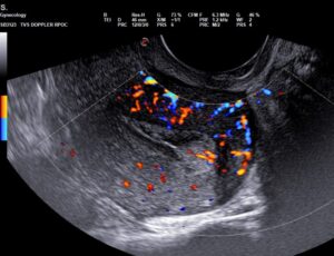
Example case:

Young lady presenting with recurrent mid cycle vaginal bleeding, advised transvaginal USG

Gynaecologic ultrasound fig.A
Endometrial polyp with increased vascularity in fundal part of endometrial cavity
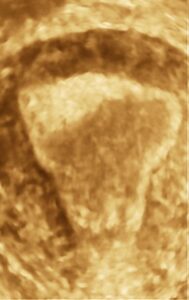
Gynaecologic ultrasound fig.B
Endometrial polyp better visualized on 3D frontal projection

What is the importance of gynecological ultrasound for women’s health?

Gynecological ultrasounds are among the most commonly performed sonographic examinations requested for women. Used as preventive measures, they contribute to the early identification of diseases and allow the treatment to start quickly, increasing the chances of cure and simplifying treatment.

pregnancy ultrasound in Pitampura

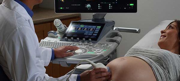
Pregnancy Ultrasound

Pregnancy Ultrasound in Pitampura: A Comprehensive Guide by Dr. Anuj Thakral

Pregnancy ultrasound is a pivotal aspect of prenatal care, offering invaluable insights into the development and well-being of the fetus. In Delhi, Dr. Anuj Thakral stands out as a leading expert for Level 1 & 2 pregnancy ultrasound in Pitampura, providing exemplary services to expectant mothers. Understanding the different levels of pregnancy ultrasound can empower individuals with the knowledge they need throughout their journey to parenthood.

Introduction to Pregnancy Ultrasound

Pregnancy ultrasound, also known as sonography, is a non-invasive imaging technique used to monitor the progression of pregnancy. By utilizing sound waves, it provides detailed images of the fetus, placenta, and uterus, allowing healthcare providers to assess fetal development and detect any potential complications.

Importance of Pregnancy Ultrasound

Ultrasound plays a crucial role in prenatal care by enabling healthcare providers to:

  • Monitor fetal growth and development.
  • Assess the placenta and amniotic fluid levels.
  • Identify any abnormalities or birth defects.
  • Determine the baby’s position and estimate its size.
  • Evaluate the health of the mother’s reproductive organs.

Why choose Dr. Thakral’s Diagnostic Centre for level 1 & 2 pregnancy ultrasound in Pitampura

When considering where to go for level 1 & 2 pregnancy ultrasounds in Pitampura, there are several reasons why Thakral’s Diagnostic Centre stands out as an excellent choice:

  1. Expertise: Dr.Thakral’s Diagnostic Centre boasts a team of highly skilled and experienced radiologists and sonographers who specialize in prenatal imaging. With their expertise, you can trust that your ultrasound will be conducted with precision and accuracy.
  2. State-of-the-art Equipment: The centre is equipped with advanced ultrasound machines that produce high-quality images, allowing for detailed assessments of fetal development at every stage of pregnancy. This ensures that any potential abnormalities or concerns can be identified early on.
  3. Comprehensive Services: Whether you need a basic ultrasound to confirm your pregnancy or a more detailed level 1 ultrasound for specialized assessment, Dr.Thakral’s Diagnostic Centre offers a full range of prenatal imaging services to meet your needs. They also provide level 2 ultrasounds for comprehensive anatomical scans.
  4. Patient-Centered Care: At Dr. Thakral’s Diagnostic Centre, patient care is paramount. The staff are dedicated to providing compassionate and personalized care to every expectant parent who walks through their doors. They understand the importance of this moment in your life and strive to make your ultrasound experience as comfortable and stress-free as possible.
  5. Convenient Location: Situated in Pitampura, Thakral Diagnostic Centre offers a convenient location for residents in the area. Whether you live nearby or are traveling from elsewhere in Delhi, you can easily access their facilities for your ultrasound appointments.
  6. Prompt and Efficient Service: Time is of the essence when it comes to prenatal care. Thakral Diagnostic Centre is known for its prompt and efficient service, ensuring that you receive your ultrasound results in a timely manner. This allows for early detection of any issues and timely interventions if necessary.

What are the types of Ultrasound studies in pregnancy  (trimester wise):

First-trimester ultrasound

Best Pregnancy Ultrasound in Pitampura

There are generally two ultrasounds offered during the first 3 months (First trimester) of pregnancy:

  1. Early pregnancy scan or viablity scan (At 6-8 weeks of pregnancy): Helps to confirm and determine pregnancy age, hence the name dating scan. This pregnancy scan can detect the baby’s heartbeats and any source of internal bleeding or abnormality found in the uterus that might affect the pregnancy.It also confirms the location of pregnancy and checks if it is inside or outside the uterus. Pregnancies outside the uterus are called ectopic pregnancies. Such pregnancies need early diagnosis and immediate medical attention. Twin pregnancy or multiple pregnancies can also be detected at this stage.

The dating scan is very important as it establishes a benchmark which will enable accurate assessment of fetal growth as the pregnancy progresses in the 2nd & 3rd trimester.

2. Nuchal Translucency Scan (NT/NB ultrasound pregnancy scan)

(11 week to 13 week 6 days ultrasound scan): Most suitable time is 12-13 weeks

“Nuchal” is the adjective for the “nape of the neck”, and translucency here refers to the translucent area in that portion. The fluid at the back of your baby’s neck is checked by this scan. The NT scan looks at the entire spinal column of the baby, measures the size and thickness of its nuchal translucency. NT is increased in up to 40% of fetuses that have a major cardiac abnormality and is associated with other structural and genetic anomalies.

This scan also checks for the presence of nasal bone.The nasal bone is ‘absent’ or hypoplastic in 50–60% of fetuses with trisomy 21.

Alongwith this sonography, a double marker blood test is recommended. This sonography and double marker blood test guides obstetricians to rule out chromosomal abnormalities like Down’s syndrome.

We routinely perform uterine artery doppler as part of NT/NB scan. This helps us prempt possiblity of increased blood pressure and related complications in the mother and growth issues in baby in later pregnancy.

Second-trimester ultrasound

 Best Pregnancy Ultrasound in Pitampura

Level 2 Pregnancy Ultrasound: A Detailed Look

Reasons for Level 2 Ultrasound

A Level 2 ultrasound, also known as an anatomy scan or targeted ultrasound, provides a more comprehensive evaluation of fetal anatomy and development. It is typically performed between 18-22 weeks of pregnancy.

What’s Examined

During a Level 2 ultrasound, Dr Anuj Thakral examines the fetus in detail, focusing on the brain, heart, spine, limbs, and other vital organs. This scan can detect structural abnormalities and assess the baby’s overall health.

When it’s Done

The timing of a Level 2 ultrasound is critical, as it allows Dr Anuj Thakral to detect any anomalies early in the pregnancy. Early detection can lead to timely interventions and better outcomes for both the mother and baby.

3. Anomaly scan/TIFFA scan (Level 2 scan)- at 18 to 20 weeks

An anomaly scan or TIFFA scan (Targeted Imaging for Fetal Anomalies) is the most essential pregnancy scan carried out in your second trimester.

The anomaly scan assesses the baby’s structural anatomy & internal organs from head to toe in adequate detail. Besides recording the fetal size & weight, a detailed anomaly scan can help in timely detection of a variety of major congenital anomalies/malformations. For this, special attention is paid to the brain, face, spine, heart, stomach, bowel, kidneys, and limbs..

During the anomaly scan, we also assess the position of placenta, the umbilical cord, and amniotic fluid around the fetus. A uterine artery doppler is also an essential part of the study.

Best Pregnancy Ultrasound in Delhi

Fetal Echocardiography

This ultrasound is meant to check the structure and function of a baby’s heart.

This is one of the optional sonographies during pregnancy. This pregnancy ultrasound scan is recommended typically in high-risk pregnancies. It’s typically done in the second trimester, between weeks 18 to 24.

This scan looks out for abnormalities such as holes in the heart, narrowing of arteries, valves that don’t open and close properly. The common indications for fetal echocardiography include:

  • Mother having a heart defect
  • Mother already has had a baby with a heart defect
  • Diabetic mother: a slightly higher risk of having a baby with a heart defect
  • History of consuming certain drugs that can increase the risk of heart problems, such as some antiepileptic medications
  • Abnormal nuchal translucency (NT) scan with normal chromosomal study

Best Pregnancy Ultrasound in Pitampura

Third-trimester ultrasound

Pregnancy Ultrasound in Pitampura

In the last 3 months of pregnancy, ultrasound is used to monitor the baby’s health, growth, development, and internal organs. Changes in the uterine environment are also carefully assessed. Following are the pregnancy ultrasound scan prescribed in the third trimester :

  1. Third Trimester growth & Fetal well being scan: besides fetal growth & anomalies, the position of placenta & amount of amniotic fluid is assessed. The lower end of the placenta should not be lying too close to the cervix. In the case of twins or triplets, there is a greater chance of developing growth problems, which is checked by the growth scan.
  2. Obstetric Doppler scan: A proper blood flow to the uterus, placenta, and the umbilical cord is crucial for a baby to develop normally inside the womb during pregnancy.

A routine pregnancy ultrasound alone is incapable of demonstrating the blood flow. It is complimented by color doppler which looks at both the maternal vessels (uterine arteries) & fetal vessels (umbilical & middle cerebral arteries in most cases). This helps identify & quantify deficencies in maternal & fetal blood flow which are important to guide further management in this final trimester.

Benefits of Early Detection

Early detection of fetal abnormalities or complications allows healthcare providers to develop a comprehensive care plan and provide appropriate support and interventions to ensure the best possible outcomes for both the mother and baby.

Risks and Safety Concerns

While ultrasound is considered safe for both the mother and fetus, it is essential to minimize unnecessary exposure to ultrasound waves. Dr Anuj Thakral follows strict guidelines to ensure the safety and well-being of both patients during ultrasound procedures.

Choosing the Right Ultrasound Facility

When selecting an ultrasound centre in Pitampura, it is essential to consider factors such as the expertise of the healthcare providers, the quality of the equipment, and the availability of comprehensive services tailored to individual needs.

Understanding the Role of Dr. Anuj Thakral

Dr. Anuj Thakral is a renowned radiologist specializing in level 1, 2, & 3 pregnancy ultrasound in Pitampura. With his expertise and compassionate care, he has helped numerous families navigate the journey of pregnancy with confidence and peace of mind.

Tips for a Successful Ultrasound Experience

  • Schedule regular prenatal appointments to monitor the progress of your pregnancy.
  • Stay hydrated and follow any specific instructions provided by your healthcare provider before the ultrasound.
  • Communicate openly with your healthcare provider about any concerns or questions you may have.
  • Take time to bond with your baby during the ultrasound by asking questions and requesting to see images of your little one.

Conclusion

Pregnancy ultrasound plays a pivotal role in prenatal care, offering invaluable insights into fetal development and well-being. By understanding the different levels of pregnancy ultrasound and the expertise of Dr Anuj Thakral, expectant parents can embark on their journey to parenthood with confidence and peace of mind.

Overall, Thakral Diagnostic Centre is a trusted choice for level 1, 2, and 3 pregnancy ultrasounds in Pitampura, offering expertise, advanced technology, comprehensive services, patient-centered care, and convenience all under one roof.

Frequently Asked Questions (FAQs)

Is ultrasound safe during pregnancy?

Yes, ultrasound is considered safe for both the mother and fetus when performed by trained healthcare professionals following established guidelines.

How often should I have a pregnancy ultrasound?

The frequency of ultrasound exams may vary depending on your specific healthcare needs and any underlying medical conditions. Your healthcare provider will determine the appropriate schedule for you.

Can ultrasound detect all birth defects?

While ultrasound is an essential tool for detecting many birth defects, it cannot identify every possible anomaly. In some cases, additional tests or screenings may be necessary.

What should I expect during a pregnancy ultrasound?

During a pregnancy ultrasound, you will lie on an examination table while a transducer is moved over your abdomen to capture images of your uterus and fetus. The procedure is painless and typically takes around 20-30 minutes.

How can I prepare for a pregnancy ultrasound?

Your healthcare provider will provide specific instructions on how to prepare for your ultrasound, which may include staying hydrated and avoiding urinating before the exam.

List of documents/information needed for any ultrasound done on a pregnant women (as per guidelines of PC/PNDT Act):

  • Identity proof of patient having full name (eg. Aadhar card) matching the prescription
  • Date of first day of last menstrual period (LMP) of pregnant lady
  • Proper prescription by referring doctor mentioning indication for ultrasound- valid only for 21 days from date of issue
  • Prescription to have full name of referring doctor
  • Prescription to have proper stamp of referring doctor with DMC number

Musculoskeletal Ultrasound

Musculoskeletal ultrasound- what is it

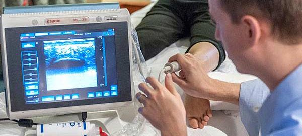
Musculoskeletal ultrasound is a non-invasive, accurate imaging modality that can prove useful in diagnosis of a variety of musculoskeletal injuries and disorders.

Advantages of musculoskeletal ultrasound over MRI include:
-dynamic nature of the study possible unlike MRI which helps provide more details pertaining to the abnormality as the scans can be performed while moving the area of interest through a range of motion.
– Comparison with the opposite side/limb is easier & retains the image quality of the area of interest. Simultaneous MR evaluation of control and affected limb is more time consuming and yet less informative with MRI
– Provides greater opportunity of clinical interaction between patient and radiologist
-Musculoskeletal interventional ultrasound can also help treat unlike MRI

A wide variety of conditions can be evaluated with musculoskeletal ultrasound:
• Tendon and ligament injuries and tears
• Lumps & bumps in relation to muscles/joints/nerves
• Joint fluid
• Peripheral nerve compression problems
• Rheumatology & Joint problems

These are some of the interesting cases that I have  recently seen in my routine practice of Musculoskeletal ultrasound:

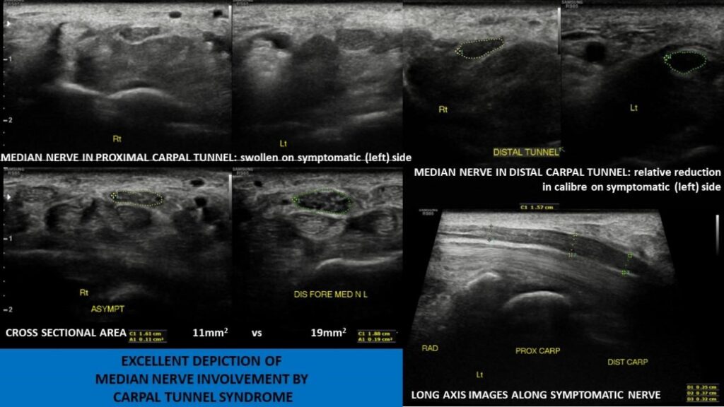
Case 1: Patient with pain in left hand with suspicion of median nerve involvement on NCV: Ultrasound clearly depicts  evidence of assymetric median nerve swelling in carpal tunnel

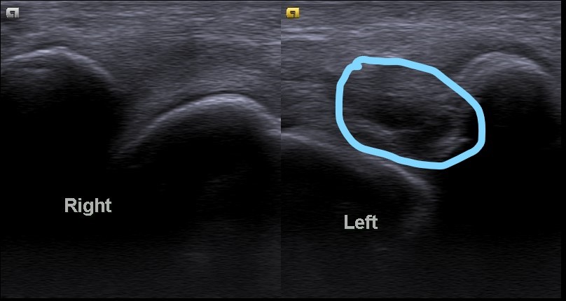
Case 2: Young female with pain and swelling in left wrist & hand: USG shows assymetric synovial thickening at left ulnocarpal joint. Diagnosed later as ankylosing spondylitis on further workup

The use of US in diagnostic and therapeutic interventional proceudres enhances the accuracy of needle placement in spaces that are difficult to enter on the basis of external landmarks alone and improves clinical outcomes.

Many USG guided interventional procedures may be performed:

  • Joint aspiration/injections
  • Synovial and mass biopsies
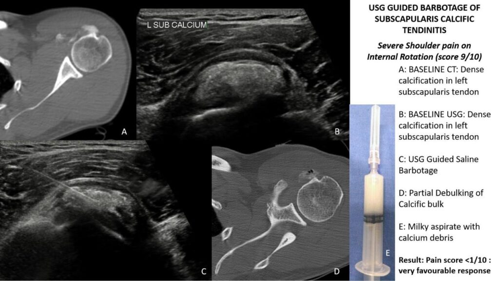
  • Barbotage for calcific tendinitis
  • Tendonsheath & Peritendoninous injections
  • Hydro-dissection techniques & Hydrodilatation for adhesive capsulitis
  • Regenerative medicine procedures including PRP & dry needling

Following are some of the interventional muscculoskeletal procedures that I have  performed recently:

Case 1: USG guided barbotage for calcific tendinitis of subscapularis muscle

Case 2: Synovial biopsy for Erosive arthritis of shoulder:

Biopsy showed presence  of granulomas suggesting tubercular etiology- patient on ATT

Case 3: Diagnostic block for confirmation of piriformis syndrome in a case of large disc bulge at L4-5 region with only unilateral symptoms. Pain disappeared after injection of local anaesthetic in piriformis muscle on symptomatic side confirming diagnosis of piriformis syndrome. Patient counselled for active stretching excercises and on follow up. Can be taken for steroid/botox injection if symptoms worsen